PRODUCTS
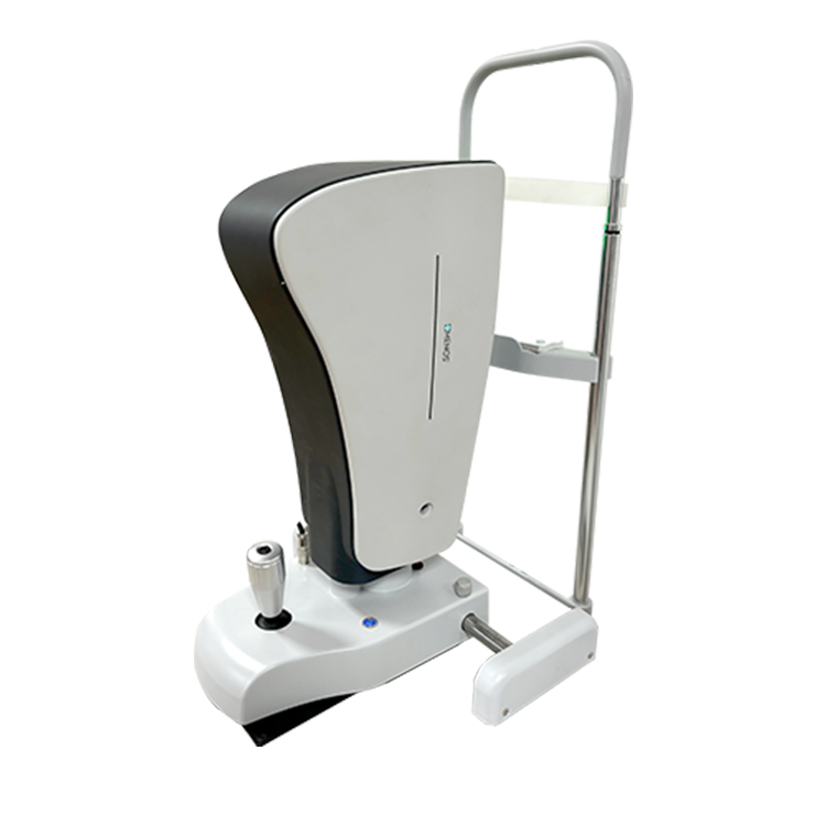
Lumino 10
OCT
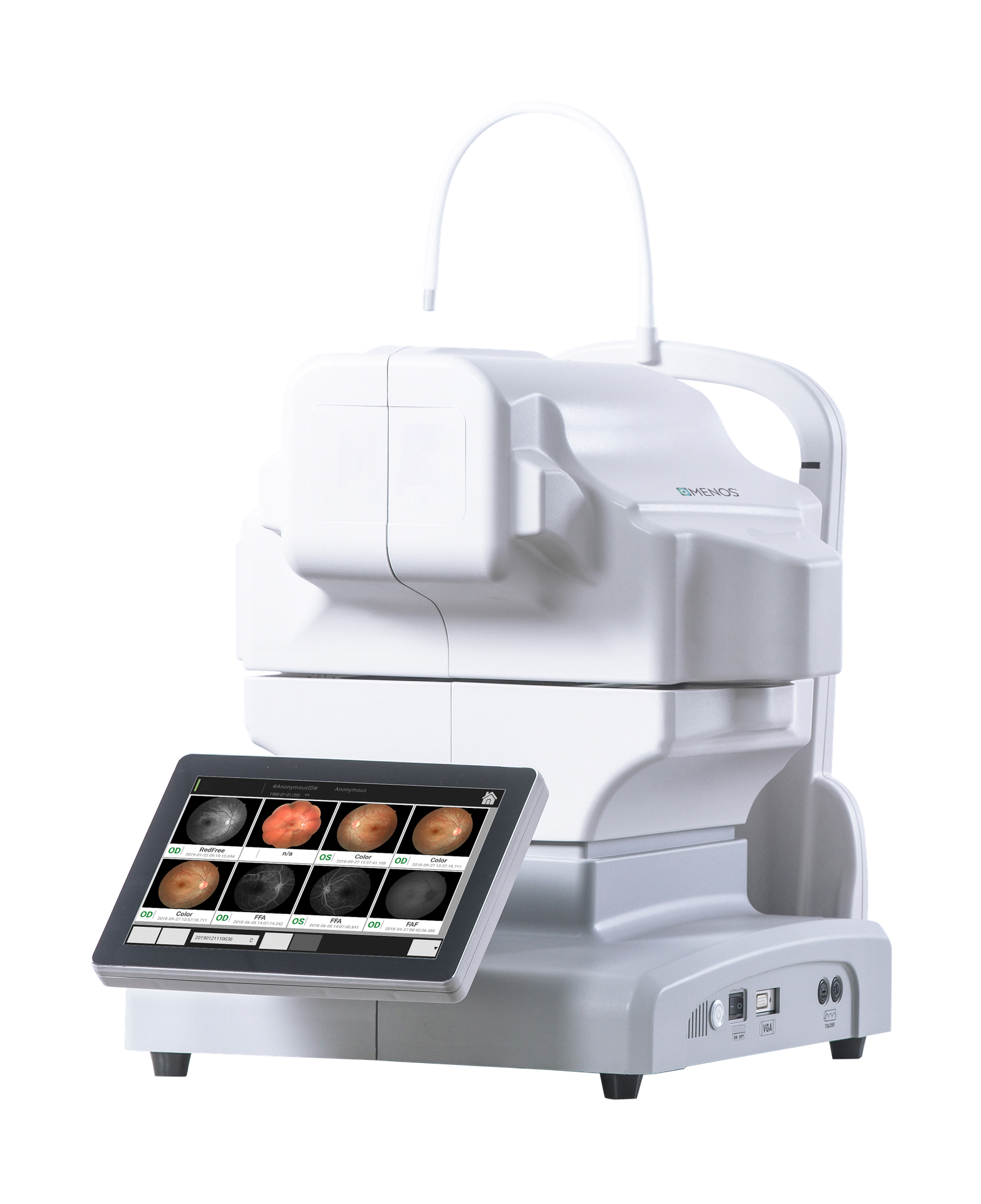
A NEW GENERATION OF FULLY AUTOMATIC ARTIFICIAL INTELLIGENCE OCT
The Lumino10 Optical Coherence Tomography for Ophthalmology is an integrated automatic artificial intelligence OCT system with both soft and hard capabilities, boasting complete independent intellectual property rights. It combines cutting-edge multi-image registration enhancement technology with industry-leading analysis techniques to offer a
comprehensive solution for clinical diagnosis and treatment. This includes automated image capture, precise image analysis, intelligent and real-time cloud-based data sharing.
ENHANCING THE EFFICACY
AND DEPENDABILITY OF
CLINICAL DIAGNOSIS

ANTERIOR SEGMENT
High definition OCT imaging of the cornea enables clear visualization of the cornea segmentation

Anterior HD single line

Anterior six Radial line
GLAUCOMA
Accurately measure and detect RNFL, TSNI and ILM-RPE to diagnosis the health to provides valuable insights
into overall retinal health
DISC AREA

SOFTWARE ANALYSIS

The macular area

SOFTWARE ANALYSIS

WIDE FIELD OCT SCAN ( 12MM X 9MM)
The Lumino1 can capture a 12mmx9mm wide field OCT scan, encompassing both the
macula and optic disc. Ideal for an annual eye exam
High definition single line scan

Six radial line scan


Diagram of the lesson

MACULAR HOLE

Macular hole is a tissue loss of the entire layer or part of the macular nerve retina. Most of them are idiopathic and related to abnormal vitreomacular traction, and a few are related to trauma. Lamellar macular hole (LMHH) is a layer of tissue loss on the surface of the fovea and is often associated with the epiretinal membrane
SOFTWARE ANALYSIS

On OCT, there was a serous detachment under the retinal neuroepithelial layer, with uniform hyporeflection within the detachment area, and a continuous light reflection zone in the retinal pigment epithelial layer below
- TECHNICAL PARAMETERS
- SCANNING MODES


